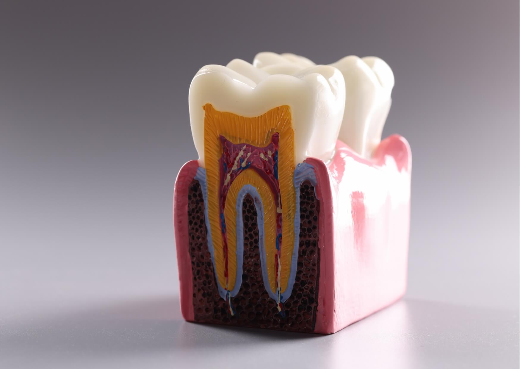
If you are wondering what a tooth nerve looks like, the short answer is that it does not look like the white outer part of a tooth.
What people call the “tooth nerve” is part of the soft tissue inside the tooth, called the pulp. When it is visible during treatment or in a badly damaged or extracted tooth, it may look like a small strand or bit of pink, red, or yellowish tissue.
Most people never actually see it. The pulp is protected by enamel, dentin, and the roots, so it stays hidden unless the tooth is badly decayed, fractured, or opened during a procedure such as a root canal.
Dulce Dental offers emergency dentistry in Dallas, TX and provides the kind of prompt care patients may need for suspected exposed nerves or severe tooth pain.
A tooth has several layers. The hard outer enamel covers the crown, dentin sits underneath, and the center holds the pulp.
The pulp contains nerve fibers, blood vessels, and connective tissue. In the crown of the tooth, this area is called the pulp chamber. In the roots, it narrows into small passageways called root canals.
So when people talk about the “tooth nerve,” they usually mean the soft tissue inside this pulp space, not a single wire-like structure running through the whole tooth.
In simple terms, a healthy tooth nerve usually looks like soft, moist tissue rather than a clean, separate cord. It may appear pale pink, reddish, or slightly yellow because it is mixed with tiny blood vessels and connective tissue.
In a small front tooth, the pulp space is narrow, so the tissue may look like a fine thread. In a larger molar, the pulp chamber is wider, so the tissue may look more like delicate soft material filling a small hollow center.
If the pulp is inflamed or infected, it may look darker red, swollen, or broken down. In a severely damaged tooth, it may not look distinct at all because the tissue can degenerate as infection progresses.
This question usually comes up when someone has sharp tooth pain, deep decay, or a broken tooth and wants to know what may be exposed. Sometimes a person sees a tiny red spot inside a damaged tooth and wonders if that is the nerve.
It might be, but not always. What looks like a “nerve” may actually be pulp tissue, blood, dentin, or debris from the damaged area. That is one reason a dental exam matters when the inside of a tooth seems visible.
A true exposed pulp can be very sensitive to cold, air, pressure, and sweets. In some cases, the pain throbs or lingers after the trigger is gone.
There is an important difference here. The tissue inside the tooth contains small nerve fibers, but those fibers connect to larger nerves in the jaw through a tiny opening at the tip of each root.
That means the “tooth nerve” is really one part of a larger nerve pathway. During root canal treatment, the tissue inside the tooth may be removed, but the gums, bone, and nearby tissues still have feeling because other nerves serve those areas.
This is why a tooth can stop having internal pulp pain after a root canal while the area around it can still feel pressure or soreness during healing.
When the pulp becomes inflamed, the condition is called pulpitis. This often happens because of deep decay, a crack, repeated dental work, trauma, or heavy wear.
Early irritation may cause brief sensitivity to cold or sweets. More advanced inflammation may cause pain that lingers, wakes someone at night, or becomes hard to pinpoint.
If the pulp loses its blood supply and dies, bacteria can spread through the root canal system into the tissues around the root. That can lead to infection, swelling, pain with biting, or an abscess.
A dental abscess needs prompt professional care. Swelling in the face, fever, trouble swallowing, or difficulty opening the mouth are urgent warning signs and should prompt immediate evaluation for a dental emergency or emergency dentistry.
Usually, no. Even if a cavity or fracture is deep, the tissue inside the tooth is small and not easy to identify accurately in a mirror.
People sometimes assume that any dark hole, red dot, or painful spot is the nerve, but that is not reliable. Teeth have several internal structures, and damage can change what is visible.
If a tooth looks broken open or has a visible inner area with significant pain, the safest next step is a dental evaluation rather than trying to inspect or test it at home.
Dentists do not diagnose pulp problems by appearance alone. They usually combine your symptoms with an exam, temperature testing, bite testing, and dental X-rays.
An X-ray does not show the nerve itself in detail, but it can show decay, deep fillings, some fractures, and changes around the root tip that suggest infection. A dentist will often order dental X-rays to check for deep decay or root involvement.
This matters because not all tooth pain comes from the pulp. Gum disease, sinus pressure, grinding, cracked teeth, and pain referred from another tooth can sometimes feel similar.
Treatment depends on how much damage the pulp has sustained. A small cavity or mild irritation may improve after the tooth is restored if the pulp is still healthy enough to recover.
In those cases, a dentist can often repair the tooth with tooth-colored fillings. If the pulp is too inflamed to heal, a dentist may recommend root canal treatment to remove the damaged tissue inside the tooth and seal the canal space.
If the tooth cannot be restored, tooth extractions may be the better option. The right choice depends on how much tooth structure remains, how deep the decay or fracture goes, the condition of the surrounding bone and gums, and the patient’s overall dental plan.

A tooth with nerve involvement may be very painful, but not always. Some teeth become less painful after the pulp dies, even though infection is starting to spread beyond the root.
That is why pain relief does not always mean the problem is gone. A tooth that changes color, develops swelling, feels high when biting, or starts draining a bad-tasting fluid needs prompt dental attention.
Persistent tooth pain is easy to underestimate. Earlier treatment is often simpler and more predictable than waiting for swelling or severe infection.
The best way to protect the tooth nerve is to treat cavities early and reduce the chance of cracks or repeated trauma. Daily brushing with fluoride toothpaste, flossing, and regular dental visits still do most of the heavy lifting.
If a tooth is sensitive for more than a short time, hurts when biting, or has a visible chip or dark area, it is worth getting checked before the pulp becomes involved. Night grinding can also stress teeth over time, so a dentist may look for wear patterns or fractures if symptoms keep returning.
If you suspect the inside of a tooth is exposed, or if pain is getting worse instead of better, a dental exam can help determine whether the pulp is irritated, infected, or no longer healthy enough to recover.
If you’re in Dallas, TX or nearby West Dallas or Oak Cliff and need emergency dentistry, Dulce Dental can help; call +1 214-337-0153 to schedule.
Not exactly, but people often use the terms as if they mean the same thing. The pulp is the soft tissue inside the tooth, and it contains nerve fibers along with blood vessels and connective tissue.
It is usually not a bright white structure like the tooth itself. It may look pink, red, yellowish, or darker if it is inflamed or breaking down.
Yes. That is the goal of root canal treatment in many cases. The dentist removes the diseased pulp tissue from inside the tooth, disinfects the internal space, and seals it so the outer tooth structure can remain in place.
It may, but not always. Blood can come from injured gum tissue or from pulp exposure, so a dental exam is the safest way to tell.
Seek urgent care if there is facial swelling, fever, trouble swallowing, or severe worsening pain. Those signs can suggest a spreading infection and should not be ignored.